The path toward a successful colonoscopy, a procedure pivotal for colorectal cancer screening and diagnosis, is often fraught with a specific kind of apprehension, which paradoxically centers less on the examination itself and more on the required preparatory phase. Patients frequently perceive the preparation as the most demanding element of the entire experience, and understanding this initial hurdle is fundamental to demystifying the whole process. The core objective of the preparation is straightforward: to fully cleanse the entire large intestine of solid matter, providing the examining physician with an unobstructed, panoramic view of the mucosal lining. Without a perfectly clean canvas, small polyps or subtle lesions can be easily obscured by residual stool, directly compromising the accuracy and, by extension, the entire preventive value of the procedure.
Patients frequently perceive the preparation as the most demanding element of the entire experience.
The preparation protocol begins not on the evening prior, but typically two to three days before the scheduled appointment, necessitating a change in dietary habits to what is broadly termed a low-residue diet. This initial phase involves systematically removing high-fiber foods that are difficult to digest and clear from the bowel, such as nuts, seeds, raw vegetables, and whole grains. The purpose is to reduce the sheer volume of undigested matter entering the colon, thereby easing the more aggressive cleansing phase that follows. Adherence to this low-residue guideline significantly influences the efficiency of the main prep, demonstrating that the success of the procedure is truly a cumulative effort beginning days in advance. Failing to respect these early dietary restrictions can lead to a less-than-optimal cleansing, which in unfortunate scenarios can result in the need to repeat the entire preparation and procedure.
The purpose is to reduce the sheer volume of undigested matter entering the colon.
The final 24 hours before the procedure constitutes the most intensive phase: the strict clear liquid diet. During this period, no solid food whatsoever is permitted. The patient’s caloric and hydration intake is limited to specific clear liquids, which may include water, clear broth, apple juice, pale-colored sports drinks, and clear gelatin, with all red, blue, or purple liquids strictly avoided as they can mimic the appearance of blood within the colon. This liquid-only diet creates a completely empty upper gastrointestinal tract, setting the stage for the powerful laxative solution. Hydration throughout this phase is critically important, not merely for comfort, but to mitigate the risk of dehydration, a genuine concern given the rapid fluid loss induced by the bowel-cleansing agents.
This liquid-only diet creates a completely empty upper gastrointestinal tract, setting the stage for the powerful laxative solution.
The actual bowel preparation solution, often a polyethylene glycol-based product, is an osmotic laxative, engineered to pull copious amounts of water into the colon, effectively flushing out its contents. This solution is generally consumed in a split-dose regimen—the first portion on the evening prior to the colonoscopy and the second portion on the morning of the procedure, a few hours before the appointment. The split-dose approach is medically preferred because it ensures the distal end of the colon, the area most frequently affected by cancer, is as clean as possible at the time of examination. The experience of drinking the solution can be challenging due to its volume and often-salty or chemical taste, and the ensuing bowel movements are predictably frequent and watery. Patients are encouraged to use strategies to improve palatability, such as chilling the solution or mixing it with approved clear liquids, and to manage potential perianal skin irritation with protective creams.
This solution is generally consumed in a split-dose regimen—the first portion on the evening prior to the colonoscopy and the second portion on the morning of the procedure.
On arrival at the endoscopy unit, the patient moves from the anxiety of the prep phase to the clinical environment. The initial steps involve necessary administrative checks, a review of the patient’s medical history, and confirmation of consent. A nurse will then prepare the patient, which includes changing into a gown and establishing an intravenous (IV) line, the conduit for hydration and, critically, for the sedative medication. This preparatory time is essential for the clinical team to ensure all safety parameters are met and to address any lingering patient questions regarding the process. The atmosphere is professional and carefully managed, minimizing unnecessary external stimulation to maintain a calm setting for the ensuing procedure.
A nurse will then prepare the patient, which includes changing into a gown and establishing an intravenous (IV) line.
The role of sedation in modern colonoscopy is paramount to patient comfort and procedural success. While the level of sedation varies—ranging from light conscious sedation to monitored anesthesia care (deep sedation), often utilizing Propofol—the goal is nearly universal: to ensure the patient feels minimal to no discomfort and has little to no memory of the actual procedure. Deep sedation, administered and monitored by an anesthesiologist or a certified registered nurse anesthetist, provides a brief, controlled period of sleep, allowing the endoscopist to focus entirely on the careful navigation and inspection of the bowel. Patients under this level of care are essentially unaware of the scope’s insertion or advancement, which significantly reduces the psychological and physical stress associated with the examination.
The role of sedation in modern colonoscopy is paramount to patient comfort and procedural success.
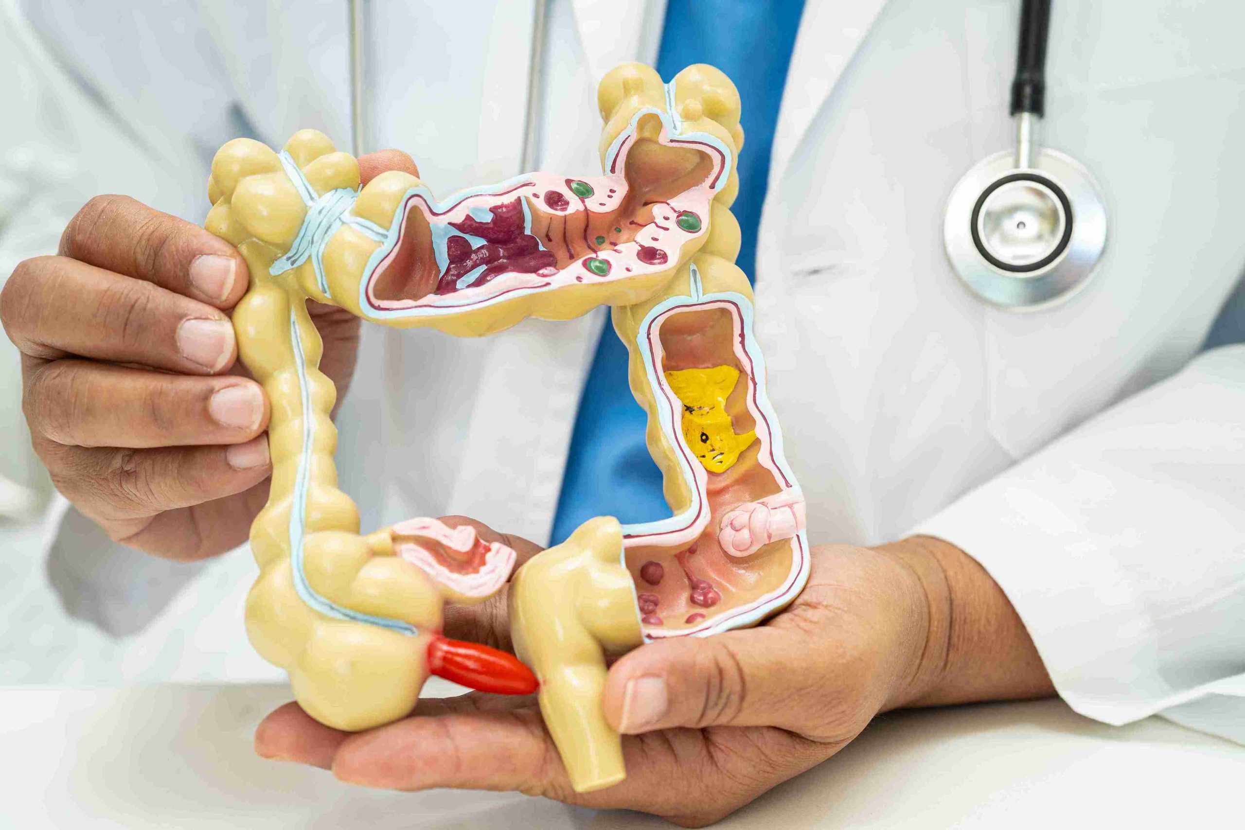
During the procedure, the patient is typically positioned on their left side. The doctor gently inserts the colonoscope, a long, flexible tube with a camera and light source at its tip, into the rectum and gradually advances it through the entire length of the large intestine to the cecum, the junction with the small intestine. To create a clear pathway and expand the intestinal walls for optimal viewing, the colonoscope insufflates the colon with air or, increasingly, with carbon dioxide. The images are relayed to a high-definition monitor, allowing the physician to meticulously inspect the colon’s inner lining. The duration of the examination is highly variable, influenced by the anatomy of the individual’s colon, which can have twists and loops, and whether any therapeutic interventions, such as polyp removal, are necessary.
The doctor gently inserts the colonoscope, a long, flexible tube with a camera and light source at its tip, into the rectum and gradually advances it through the entire length of the large intestine to the cecum.
Polyp identification and removal constitute the central therapeutic benefit of the colonoscopy. Polyps are abnormal growths on the lining of the colon, many of which are benign but possess the potential to evolve into malignant tumors over time. If the physician identifies a polyp, it is typically removed immediately during the procedure using specialized instruments passed through the scope, a process called polypectomy. Small pieces of tissue can also be collected for laboratory analysis (biopsy) if suspicious areas are noted. This dual diagnostic and therapeutic capability is what makes the colonoscopy the gold standard for colorectal cancer prevention, as the removal of pre-cancerous tissue halts the progression of the disease.
Polyp identification and removal constitute the central therapeutic benefit of the colonoscopy.
Immediately after the scope is withdrawn, the patient is transferred to a recovery area. The effects of the sedation dissipate relatively quickly, though a period of monitoring is essential until the patient is fully alert and their vital signs are stable. The most common immediate sensation experienced in recovery is a feeling of abdominal fullness, bloating, and the need to pass gas. This is directly attributable to the insufflated air or carbon dioxide remaining in the colon. Nurses actively encourage patients to walk or move, as expelling this residual gas provides significant and rapid relief from the cramping. Upon discharge, which usually occurs within an hour or two, patients are strictly prohibited from driving and must be escorted home, a non-negotiable safety requirement due to the lingering effects of the sedative.
The most common immediate sensation experienced in recovery is a feeling of abdominal fullness, bloating, and the need to pass gas.
The instructions for the first 24 hours of home recovery are straightforward, focusing primarily on rest, hydration, and a gradual reintroduction of food. Patients are advised to avoid alcohol, strenuous activity, and any tasks requiring fine motor skills or judgment. A light, easily digestible diet is recommended for the initial post-procedure meal. Although the procedure is considered extremely safe, patients are educated to monitor for a few, rare warning signs, such as severe, persistent abdominal pain, heavy rectal bleeding, or fever, which would necessitate immediate contact with the care team. The results of the examination are often communicated to the patient shortly after the procedure, with pathology results for any removed polyps following within a few weeks, completing the patient journey from preparation to definitive findings.
A light, easily digestible diet is recommended for the initial post-procedure meal.
A colonoscopy is a multi-stage process where stringent preparation, not the brief, sedated procedure, is the primary key to achieving life-saving diagnostic clarity.